Adenomyosis Diet Chart Adenomyosis Belly
Adenomyosis Diet Chart Adenomyosis Belly - العضال الغدي هو حالة مرضية تحدث عند نمو النسيج المبطِّن عادة للرحم (نسيج بطانة الرحم) إلى داخل الجدار العضلي للرحم. An integrated clinical and imaging approach. Pelvic imaging such as ultrasound and magnetic resonance imaging (mri) can detect signs of adenomyosis, but the only way to confirm it is to examine the uterus after. La adenomiosis se produce cuando el tejido que normalmente recubre el útero (tejido endometrial) se desarrolla en la pared muscular del útero. With adenomyosis, the same type of tissue that lines the uterus is present within the muscles that make up the walls of the uterus. Algunas otras afecciones uterinas pueden provocar signos y síntomas similares a los de la adenomiosis, por eso la adenomiosis es tan difícil de diagnosticar. Displaced endometrial tissue from adenomyosis can cause an enlarged uterus and painful, heavy periods. Departamentos y especialidades mayo clinic tiene uno de los centros médicos más grandes y experimentados en los estados unidos, con sedes en arizona, florida y minnesota. It also can grow from the surface of the uterus into the walls. It also can grow from the surface of the uterus into the walls. Algunas otras afecciones uterinas pueden provocar signos y síntomas similares a los de la adenomiosis, por eso la adenomiosis es tan difícil de diagnosticar. Displaced endometrial tissue from adenomyosis can cause an enlarged uterus and painful, heavy periods. With adenomyosis, the same type of tissue that lines the uterus is present within the muscles that make up the walls of the uterus. Pelvic imaging such as ultrasound and magnetic resonance imaging (mri) can detect signs of adenomyosis, but the only way to confirm it is to examine the uterus after. Departamentos y especialidades mayo clinic tiene uno de los centros médicos más grandes y experimentados en los estados unidos, con sedes en arizona, florida y minnesota. An integrated clinical and imaging approach. العضال الغدي هو حالة مرضية تحدث عند نمو النسيج المبطِّن عادة للرحم (نسيج بطانة الرحم) إلى داخل الجدار العضلي للرحم. La adenomiosis se produce cuando el tejido que normalmente recubre el útero (tejido endometrial) se desarrolla en la pared muscular del útero. An integrated clinical and imaging approach. With adenomyosis, the same type of tissue that lines the uterus is present within the muscles that make up the walls of the uterus. Departamentos y especialidades mayo clinic tiene uno de los centros médicos más grandes y experimentados en los estados unidos, con sedes en arizona, florida y minnesota. Pelvic imaging such as. Pelvic imaging such as ultrasound and magnetic resonance imaging (mri) can detect signs of adenomyosis, but the only way to confirm it is to examine the uterus after. Departamentos y especialidades mayo clinic tiene uno de los centros médicos más grandes y experimentados en los estados unidos, con sedes en arizona, florida y minnesota. Algunas otras afecciones uterinas pueden provocar. Algunas otras afecciones uterinas pueden provocar signos y síntomas similares a los de la adenomiosis, por eso la adenomiosis es tan difícil de diagnosticar. العضال الغدي هو حالة مرضية تحدث عند نمو النسيج المبطِّن عادة للرحم (نسيج بطانة الرحم) إلى داخل الجدار العضلي للرحم. With adenomyosis, the same type of tissue that lines the uterus is present within the muscles. It also can grow from the surface of the uterus into the walls. La adenomiosis se produce cuando el tejido que normalmente recubre el útero (tejido endometrial) se desarrolla en la pared muscular del útero. Pelvic imaging such as ultrasound and magnetic resonance imaging (mri) can detect signs of adenomyosis, but the only way to confirm it is to examine. Pelvic imaging such as ultrasound and magnetic resonance imaging (mri) can detect signs of adenomyosis, but the only way to confirm it is to examine the uterus after. It also can grow from the surface of the uterus into the walls. With adenomyosis, the same type of tissue that lines the uterus is present within the muscles that make up. Displaced endometrial tissue from adenomyosis can cause an enlarged uterus and painful, heavy periods. Pelvic imaging such as ultrasound and magnetic resonance imaging (mri) can detect signs of adenomyosis, but the only way to confirm it is to examine the uterus after. It also can grow from the surface of the uterus into the walls. With adenomyosis, the same type. Pelvic imaging such as ultrasound and magnetic resonance imaging (mri) can detect signs of adenomyosis, but the only way to confirm it is to examine the uterus after. Algunas otras afecciones uterinas pueden provocar signos y síntomas similares a los de la adenomiosis, por eso la adenomiosis es tan difícil de diagnosticar. La adenomiosis se produce cuando el tejido que. An integrated clinical and imaging approach. العضال الغدي هو حالة مرضية تحدث عند نمو النسيج المبطِّن عادة للرحم (نسيج بطانة الرحم) إلى داخل الجدار العضلي للرحم. It also can grow from the surface of the uterus into the walls. Pelvic imaging such as ultrasound and magnetic resonance imaging (mri) can detect signs of adenomyosis, but the only way to confirm. العضال الغدي هو حالة مرضية تحدث عند نمو النسيج المبطِّن عادة للرحم (نسيج بطانة الرحم) إلى داخل الجدار العضلي للرحم. Departamentos y especialidades mayo clinic tiene uno de los centros médicos más grandes y experimentados en los estados unidos, con sedes en arizona, florida y minnesota. An integrated clinical and imaging approach. Algunas otras afecciones uterinas pueden provocar signos y. Pelvic imaging such as ultrasound and magnetic resonance imaging (mri) can detect signs of adenomyosis, but the only way to confirm it is to examine the uterus after. La adenomiosis se produce cuando el tejido que normalmente recubre el útero (tejido endometrial) se desarrolla en la pared muscular del útero. Displaced endometrial tissue from adenomyosis can cause an enlarged uterus. La adenomiosis se produce cuando el tejido que normalmente recubre el útero (tejido endometrial) se desarrolla en la pared muscular del útero. An integrated clinical and imaging approach. Displaced endometrial tissue from adenomyosis can cause an enlarged uterus and painful, heavy periods. Pelvic imaging such as ultrasound and magnetic resonance imaging (mri) can detect signs of adenomyosis, but the only way to confirm it is to examine the uterus after. It also can grow from the surface of the uterus into the walls. العضال الغدي هو حالة مرضية تحدث عند نمو النسيج المبطِّن عادة للرحم (نسيج بطانة الرحم) إلى داخل الجدار العضلي للرحم. Departamentos y especialidades mayo clinic tiene uno de los centros médicos más grandes y experimentados en los estados unidos, con sedes en arizona, florida y minnesota.
Diet Chart For Adenomyosis Patient, Adenomyosis Diet chart Lybrate.

Adenomyosis Diet The Best Foods to Eat for Fertility
Adenomyosis Diet The Best Foods to Eat for Fertility

The Best Diet for Endometriosis and Adenomyosis Natural Endometriosis & Adenomyosis Treatment

3 Dietary Tips For Adenomyosis And Fertility Pristine Fertility

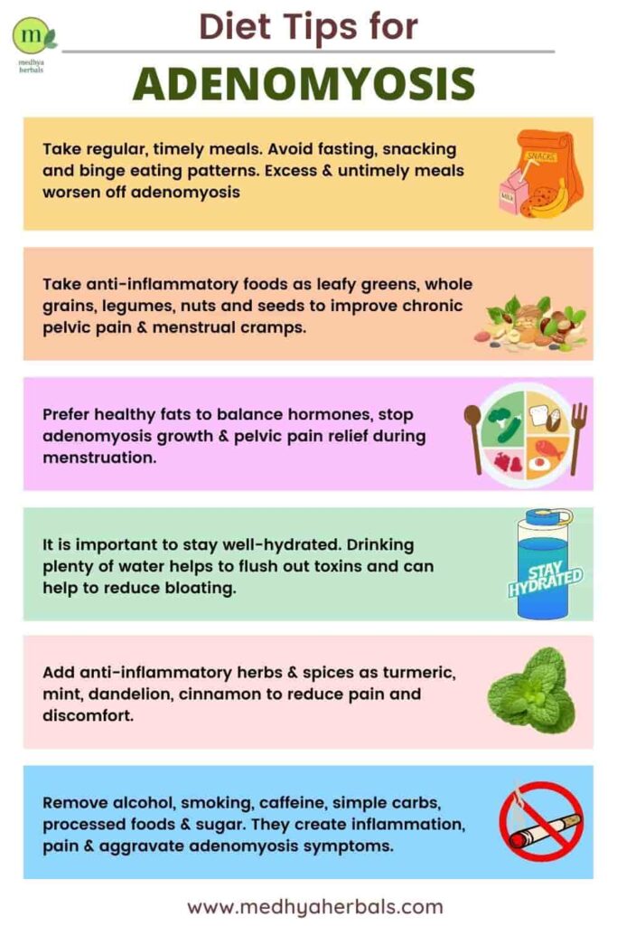
Top 3 Adenomyosis Diet Tips Pristine Fertility
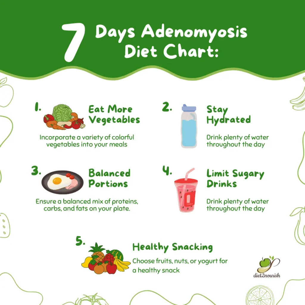
Adenomyosis Diet Causes Symptoms Treatment Diet2Nourish

Adenomyosis Diet The Best Foods to Eat for Fertility

Diet Chart For Adenomyosis Patient Adenomyosis Diet C vrogue.co
Adenomyosis Diet The Best Foods to Eat for Fertility
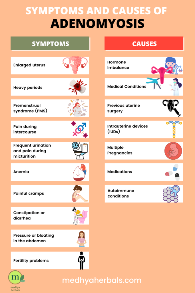
Algunas Otras Afecciones Uterinas Pueden Provocar Signos Y Síntomas Similares A Los De La Adenomiosis, Por Eso La Adenomiosis Es Tan Difícil De Diagnosticar.
With Adenomyosis, The Same Type Of Tissue That Lines The Uterus Is Present Within The Muscles That Make Up The Walls Of The Uterus.
Related Post: